Article Review: Assessing non invasive quantitative methods for 18F SynVesT 1 PET imaging of synaptic vesicle glycoprotein 2A in the rat brain
Objectives
- Validated quantitative kinetic modeling methods for [18F]SynVesT-1 PET imaging in rats.
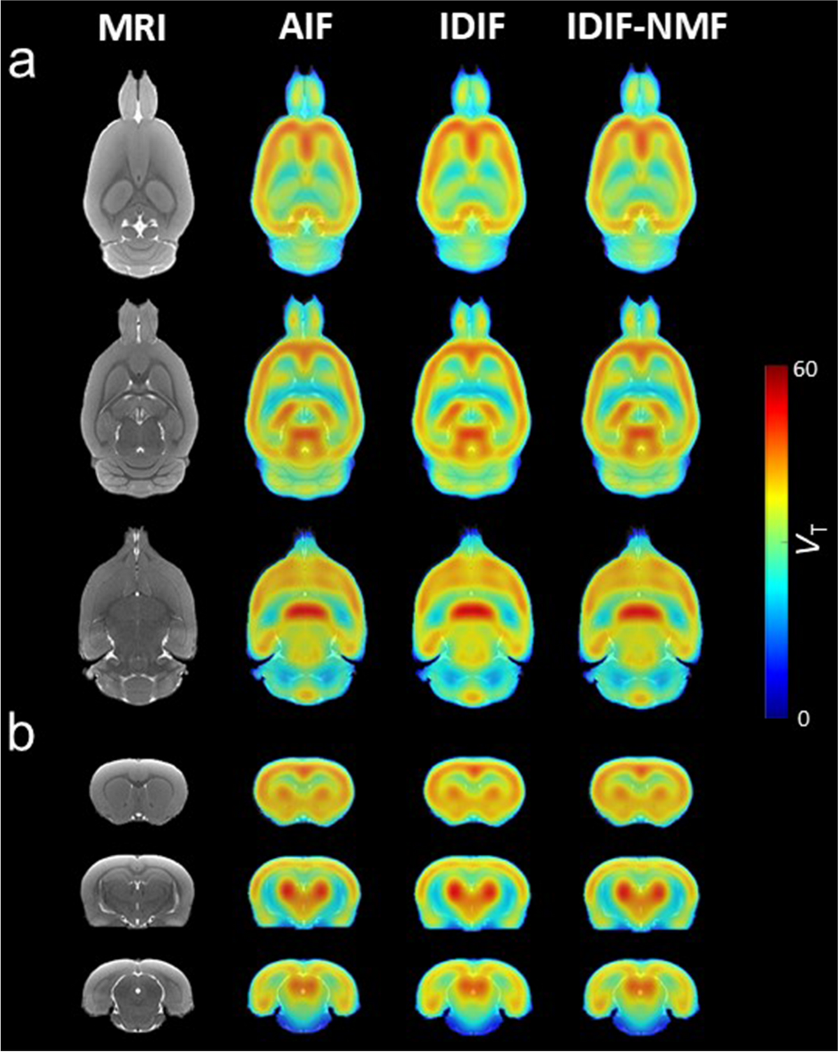
- Developed non-invasive image-derived input functions (IDIF and IDIF-NMF) as reliable alternatives to arterial input function (AIF) for quantifying synaptic density.
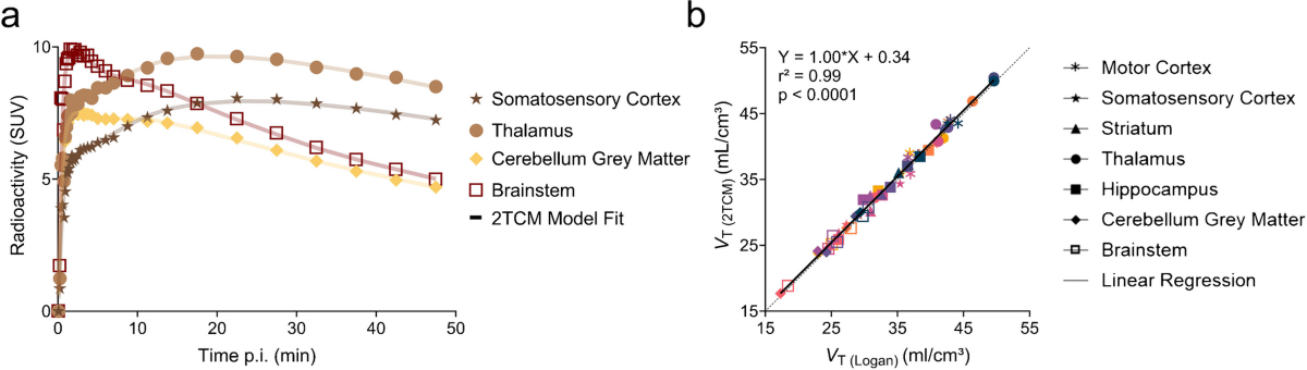
- Demonstrated that the two-tissue compartmental model (2TCM) and Logan plot are accurate methods for quantification of [18F]SynVesT-1 kinetics.
Methodology
- Used compartmental analysis approaches (1TCM, 2TCM, 3TCM) and Logan plot for kinetic modeling.
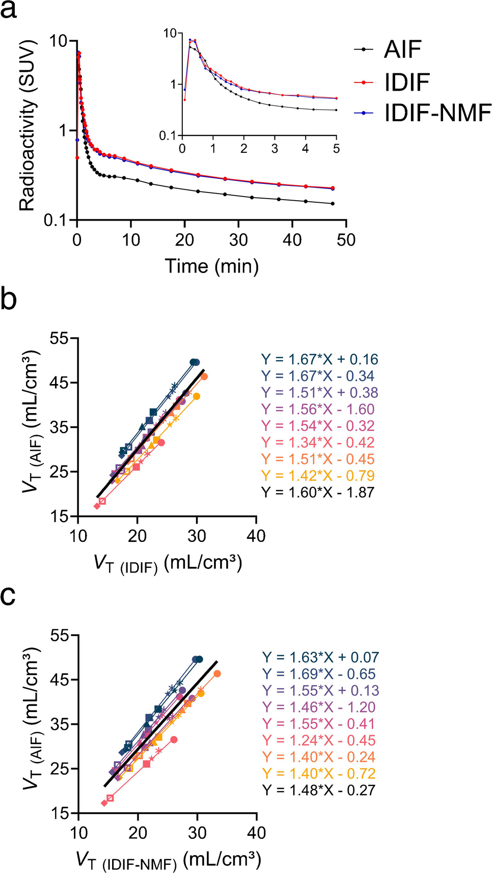
- Compared image-derived input functions (IDIF), with and without non-negative matrix factorization (NMF), against the arterial input function (AIF).
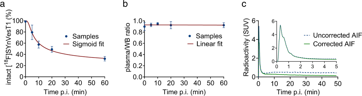
- Performed blood analysis of [18F]SynVesT-1 to generate metabolite-corrected plasma input functions.
- Utilized a sigmoid curve to fit the parent fraction of the tracer over time.
Results
- The parent fraction of [18F]SynVesT-1 decreased over time, with 48.9 ± 4.8% remaining at 20 min post-injection and 32.2 ± 3.6% at 60 min.
- The plasma-to-whole blood ratio remained stable over time (0.89 ± 0.02).
- 2TCM and Logan plot showed strong agreement (slope = 1.00, r2 = 0.99, p < 0.0001).
- Strong agreement between AIF-derived and image-derived VT values (IDIF: slope = 1.60, r2 = 0.99, p < 0.0001; IDIF-NMF: slope = 1.48, r2 = 0.99, p < 0.0001).
Discussions
- The study is well-designed and addresses an important methodological gap in preclinical PET imaging of synaptic density.
- The use of a population-based metabolite correction curve is a limitation, although justified by the low inter-animal variability. Future studies could explore individual animal correction if feasible.
- While both IDIF and IDIF-NMF performed similarly, the authors correctly point out the advantages of IDIF-NMF in terms of automation and user-independence. This should be emphasized more strongly.
- The comparison with SUV is useful, but the limitations of SUV in pathological conditions should be discussed more explicitly.