Article Review: Subendocardial quantification enhances coronary artery disease detection in 18F flurpiridaz PET
Objectives
- Developed an automated approach for evaluating subendocardial analysis for stress total perfusion deficit (TPD) and ischemic TPD using 18F-flurpiridaz PET myocardial perfusion imaging (MPI).
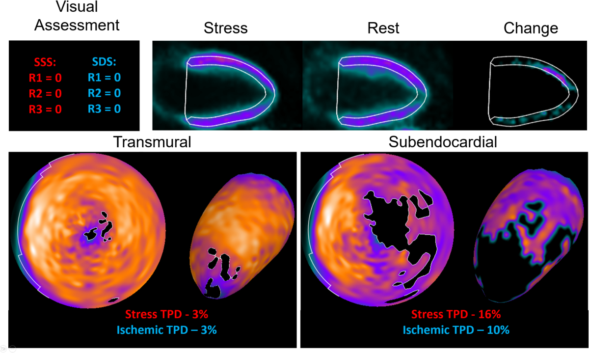
- Demonstrated that subendocardial analysis improves the detection of coronary artery disease (CAD) compared to transmural quantitative analysis or expert visual reading, especially for moderate (≥50%) stenosis.
- Showed that subendocardial and transmural TPD achieved diagnostic performance comparable to or greater than that of the readers' assessments across different subgroups.
Methodology
- Retrospective analysis of data from the 18F-flurpiridaz phase III clinical trial (flurpiridaz-301, NCT01347710).
- Automated definition of the subendocardial layer on left ventricular contours.
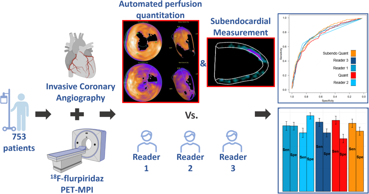
- Derivation of polar maps for both subendocardial and transmural analysis.
- Quantification of stress and rest TPD using QPET software, with ischemic TPD calculated as the difference between stress and rest TPD.
- Development of normal databases using 15 patients with visually normal studies from the phase II 18F-flurpiridaz trial.
Results
- In 753 cases, the area under the receiver operating characteristic curve (AUC) for detecting ≥50% stenosis was significantly higher for subendocardial than transmural analysis for stress (0.795 vs. 0.762, p=0.013) and ischemic (0.795 vs. 0.767, p=0.049) TPD.
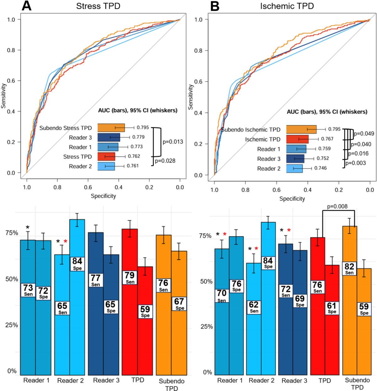
- Subendocardial TPD achieved diagnostic performance greater than or comparable to readers' assessments in the total population and across subgroups (sex, stress type, BMI).
- For ≥70% stenosis, AUC results for subendocardial and transmural quantitation were similar (p=0.339 for stress TPD, p=0.978 for ischemic TPD).
Discussions
- The study demonstrates a novel and potentially clinically relevant application of subendocardial analysis in 18F-flurpiridaz PET MPI. However, the reliance on invasive coronary angiography (ICA) as the reference standard, rather than a functional measure like fractional flow reserve (FFR), is a limitation.
- While the automated quantification is promising, the need for manual correction of contours in a small percentage of cases (2.4% stress, 4.3% rest) suggests further refinement is needed for full automation.
- The study lacks test-retest variability data, which is crucial for assessing the reliability of the new technique.
- The normal database was developed using only 15 patients. A larger and more diverse normal database would strengthen the generalizability of the findings.
Reference: Subendocardial quantification enhances coronary artery disease detection in 18F flurpiridaz PET