Article Review: Clinical benefits of LAFOV PET CT in growing demand for molecular imaging
Objectives
- The authors describe their experience transitioning from standard axial field of view (SAFOV) PET/CT scanners to a long axial field of view (LAFOV) PET/CT scanner (Siemens Biograph Vision Quadra) for routine clinical imaging in a tertiary cancer center.
- They highlight the benefits of LAFOV PET/CT, including increased patient throughput, improved image quality, reduced radiation dose, and lower per-scan costs.
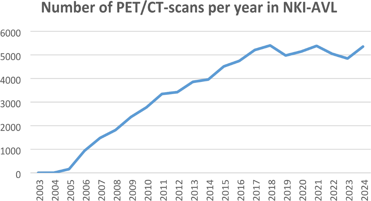
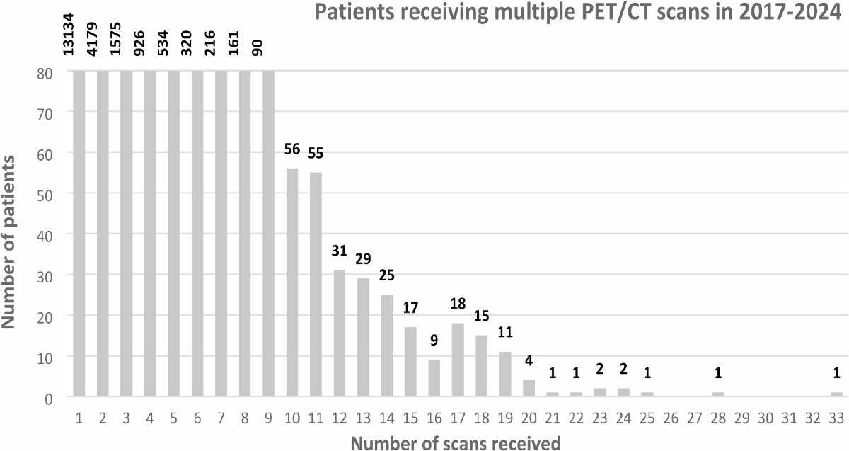
- They demonstrate the increasing demand for PET/CT scans and the growing number of patients receiving multiple scans over time.
Methodology
- The authors switched from two SAFOV PET/CT scanners to one LAFOV PET/CT scanner.
- SAFOV scanners: [18F]FDG dose of 3.5 MBq/kg, 20-30 min emission acquisition time, 30-45 min time slots.
- LAFOV scanner: [18F]FDG dose of 1.5 MBq/kg, 6 min emission acquisition time, 15 min time slots.
- The LAFOV scanner allows for imaging of all major organs in one bed position, with higher sensitivity and spatial resolution.
Results
- The transition to LAFOV PET/CT allowed for an increase in potential patient throughput from 20-25 scans per day to 32 scans per day (or more with shorter time slots).
- The administered [18F]FDG dose was reduced from 3.5 MBq/kg to 1.5 MBq/kg.
- The average radiation dose was reduced from 4.7 mSv to 2.0 mSv for a 70 kg male (or from 7.2 to 4.5 mSv, -37%, including a 2.5 mSv low-dose CT).
- Between 2017-2024, 21,414 patients underwent 41,872 scans, averaging about 2 scans per patient. 38.7% of patients received more than one scan.
Discussions
- The authors present a compelling case for the adoption of LAFOV PET/CT in high-volume clinical settings, but the report is primarily based on a single-center experience. A more rigorous comparison, perhaps with a matched cohort from the SAFOV era, would strengthen the conclusions.
- While cost savings are mentioned, a detailed cost-benefit analysis, including the initial investment, maintenance, and operational costs, would be beneficial.
- The study lacks a direct comparison of image quality between SAFOV and LAFOV, relying instead on reported improvements. Quantitative image quality metrics (e.g., standardized uptake value (SUV) measurements, lesion detectability) would provide more objective evidence.
Reference: Clinical benefits of LAFOV PET CT in growing demand for molecular imaging
3 views